Research Article Open Access
Applications of Rat Brain Synaptic Vesicle Proteins for Sensitive and Specific Detection of Botulinum Neurotoxins
Manglesh Kumar Singh, Vinita Chauhan and Ram Kumar Dhaked*
Biotechnology Division, Defence Research & Development Establishment, Gwalior-474002, India
- *Corresponding Author:
- Ram Kumar Dhaked
Biotechnology Division
Defence Research & Development Establishment
Gwalior-474002, MP, India
Tel: 91-751-2233489
Fax: 91-751-2341148
E-mail: ramkumardhaked@hotmail.com
Received Date: April 09, 2012; Accepted Date: May 16, 2012; Published Date: May 23, 2012
Citation: Singh MK, Chauhan V, Dhaked RK (2012) Applications of Rat Brain Synaptic Vesicle Proteins for Sensitive and Specific Detection of Botulinum Neurotoxins. J Bioterr Biodef S2:008. doi:10.4172/2157-2526.S2-008
Copyright: © 2012 Singh MK, et al. This is an open-access article distributed under the terms of the Creative Commons Attribution License, which permits unrestricted use, distribution, and reproduction in any medium, provided the original author and source are credited.
Visit for more related articles at Journal of Bioterrorism & Biodefense
Abstract
We propose here the application of synaptic vesicle proteins isolated from rat brain as a sole substrate for the specific endoproteinase activities of all seven serotypes of Botulinum Neurotoxin (BoNT/A to G). In this study, we used these proteins for evaluating endopeptidase and receptor binding activity for detecting BoNT/A by western blot and surface plasmon resonance with 6.25 pM and 0.22 fM limit of detection, respectively. Substrate and receptor present in the synaptic vesicle proteins are very robust and stable for more than 6 months to use in BoNT detection.
Keywords
Botulinum neurotoxin; Endopeptidase; Synaptic vesicle; SNAP-25, SPR
Background
Botulinum Neurotoxins (BoNTs) are causative agents of botulism and because of their extreme potency, lethality, ease of production, transport and the need for prolonged medical care they, are classified as category A (the highest priority) bioterrorism agents/diseases by the Centers for Disease Control and Prevention [1]. Seven distinct serotypes of the BoNTs have been identified and are designated type A through G. Humans are usually exposed to the preformed neurotoxins produced by Clostridium botulinum through food poisoning, although there are rare incidents of wound botulism and colonizing infections in the small intestine of neonates known as infant botulism [2]. Once ingested or inhaled into the lung, BoNTs are taken up by the blood stream, target the peripheral cholinergic nerve endings resulting in flaccid muscle paralysis, which leads to death. In recent years, the development of in vitro assays for the detection of BoNTs has been accelerated with the aim of decreasing the Limit Of Detection (LOD) upto femtogram quantities. However, none of the assays developed to date has been validated to be robust enough to completely replace the mouse bioassay, the FDA-approved and widely accepted gold standard method for BoNT detection. The endopeptidase assays and its variants viz. fluorescence endopeptidase assays, Fluorescence Resonance Energy Transfer (FRET) assays which can detect the biologically active neurotoxin, are reported to be more sensitive than the mouse assay [3] and have the potential to replace the mouse lethality assay. Detection of the cleaved peptides by mass spectrometry further increases the sensitivity [4], but due to the requirement for expensive instrumentation, reagents and specialized skills, these techniques are not easily affordable for most laboratories. In the scenario of a bioterror attack, an economic technology compatible with a hand-held type of device would be preferred solution for field deployment and laboratory detection.
Introduction
BoNTs act on the SNARE [soluble NSF (N-ethylmaleimide sensitive factor) attachment receptor] proteins and hydrolyze the peptide bond in a specific location resulting in inhibition of acetylcholine release. Vesicle-Associated Membrane Protein 2 (VAMP-2) is cleaved by BoNT/B, D, F and G; Synaptosomal-Associated Protein 25 (SNAP- 25) is cleaved by BoNT/A, C, and E; and syntaxin is an exclusive substrate for BoNT/C [3]. VAMP-2, SNAP-25 and syntaxin are all synaptic vesicle (SV) proteins that can be isolated from the rat brain. This indicates that synaptic vesicle proteins isolated from rat brain can be used as a substrate for detection of all the serotypes of BoNTs. The cleaved fragments of these proteins can be detected by western blot using specific antibodies indicative of BoNT serotype. The assay can differentiate between active and inactive toxin which is generally difficult for other immunoassays. The assay can also be performed in hrs compared with mouse bioassay that requires several days for serotype detection. Antibodies against SNAP-25, Syntaxin, and VAMP-2 are available from Sigma-Aldrich, Santa Cruz Biotechnology Inc., CRP Inc., Pierce Biotechnology Inc., USA. Those serotypes that exhibit highest sequence similarity share the same protein receptor, e.g., BoNT /A, E, and F bind to SV2, whereas Serotypes B and G bind to synaptotagmin I and II [5]. We herein report the application of the synaptic vesicle proteins from the rat brain as a substrate for detecting the endoproteinase activity of the botulinum neurotoxin. The binding affinity of BoNT/A heavy chain to its protein receptor present on rat brain synaptosome has also been exploited for the development of a specific and sensitive detection system for botulinum neurotoxin type A.
Methods/Discussion
Rat was anesthetized by halothane inhalation, brain was removed immediately and rinsed in ice cold 0.9 % NaCl. Fresh rat brain was homogenized with a teflon homogenizer in ten times volume (w/v) of cold homogenization buffer (25 mM Tris, 100 mM NaCl, 19.2 mM glycine, 100 μg/ml BSA, 0.1 mM DTT, 10 μM ZnCl2, pH 7.5). Homogenized samples were centrifuged at 10,000 xg for 20 min and the supernatant was collected. SV proteins present in the supernatant were then filtered through a 0.22 μ membrane filter and aliquots were stored at -20°C. The synaptic vesicle proteins obtained from a single rat brain were ~20 ml with a concentration of 2.5 mg/ml which would be sufficient for millions of detection and binding experiments and were found to be stable for more than 6 months at -20°C. The animal experiments were approved by the Laboratory Ethical Committee on Animal Experimentation of Defence Research Development Establishment (DRDE), Gwalior, India via permission no. BT/01// DRDE/ 2009.
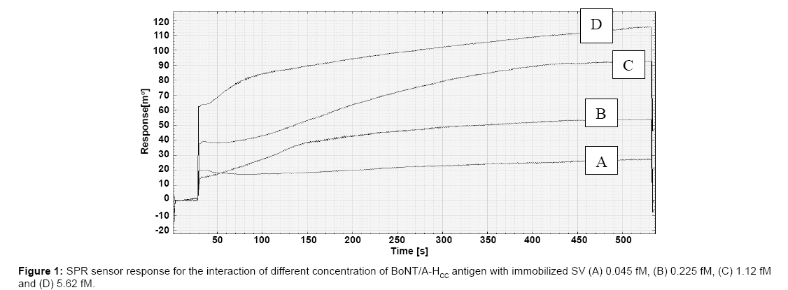
To evaluate the functional state of the isolated protein (rBoNT/A), binding of recombinant C-terminal heavy chain fusion protein (rBoNT/ A-LC-HCC) was determined by ELISA as reported in our earlier study [6] in which it was also observed that denatured rBoNT/A-LC-HCC was not able to bind with its receptor. Surface plasmon resonance (SPR) spectroscopy allows real-time measurement of interactions between immobilized ligands such as antibodies and analytes in solution. Labelling of molecules is not required and automatic analysis of small samples at low nanomolar concentrations can be performed [7]. Various SPR assays were reported for the measurement of binding of BoNT/A to polysialoganglioside GT1b [8], BoNT/A to monoclonal antibodies [9] and protein chip membrane capture assay [10]. The synaptic vesicle proteins were immobilized on a sensor chip and utilized for detection of different concentrations of C-terminal binding domain of heavy chain (rBoNT/A-HCC) resulting in a typical sensogram as depicted in figure 1. This sensogram shows concentration dependant resonance angle changes, upon the interaction of various concentrations of rBoNT/A-HCC with the immobilized proteins. Increased rBoNT/AH CC concentration (from 0.045 to 5.6 fM) resulted in increased resonance. The affinity interaction between receptor protein and rBoNT/A-HCC was characterized by the equilibrium constant (KD) and was found to be 0.22 fM using kinetic evaluation software version 5.1 (Ecochemie B.V., The Netherlands). This low KD value clearly indicates high affinity interaction of the synaptic vesicle proteins with binding domain of rBoNT/A-HCC (KD ≤ 10 nM represents the high affinity interactions) [11]. This high affinity interaction is very useful for detection of botulinum neurotoxin as low affinity antibodies limits antigen detection of such agents in low nanomolar range [12]. The binding of the receptor present in SV proteins with C-terminal domain of BoNTs can also be exploited to capture/concentrate toxin for further detection by endopeptidase assay.
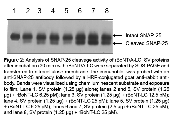
The purified recombinant BoNT/A light chain (rBoNT/A-LC) stored in 20 mM HEPES buffer (10 % glycerol, 200 mM NaCl, pH 7.5) was used for endopeptidase assay. The cleavage reaction was optimized with respect to concentrations of substrate (SV proteins, 40- 0.25 μg) and rBoNT/A-LC (1 μM to 6.25 pM), reaction incubation time (5 min to 12 hrs) and composition of cleavage buffers (data not shown). Catalytic activity of rBoNT/A-LC protein was performed at 37°C in 40 μl reaction mixture in homogenization buffer. The optimum detectable activity was recorded with 2.5 μg substrate and 12.5 pM rBoNT/ALC concentrations however; the LOD for rBoNT/A-LC was 6.25 pM at 1.25 μg substrate (Figure 2). Comparatively less intense bands were observed when lower concentrations of rBoNT/A-LC and/or substrate were used. In our observation, protein substrate is very robust, stable upon long term storage and can be used to detect endopeptidase activity of botulinum neurotoxin type A (upto 12 months, data not shown). The receptor present in the synaptic vesicle protein preparation was also found to bind with C-terminal binding domain of BoNT/A after long term storage.
Figure 2: Analysis of SNAP-25 cleavage activity of rBoNT/A-LC. SV proteins after incubation (30 min) with rBoNT/A-LC were separated by SDS-PAGE and transferred to nitrocellulose membrane, the immunoblot was probed with an anti-SNAP-25 antibody followed by a HRP-conjugated goat anti-rabbit antibody. Bands were visualized using chemiluminescent substrate and exposure to film. Lane 1, SV protein (1.25 μg) alone; lanes 2 and 5, SV protein (1.25 μg) + rBoNT-LC 6.25 pM); lane 3, SV protein (1.25 μg) + rBoNT-LC 12.5 pM); lane 4, SV protein (1.25 μg) + rBoNT-LC 25 pM); lane 5, SV protein (1.25 μg) + rBoNT-LC 6.25 pM); lanes 6 and 7, SV protein (2.5 μg) + rBoNT-LC 25 pM); and lane 8, SV protein (1.25 μg) + rBoNT-LC 25 pM).
Conclusion
The work presented here reasonably describes the technical validity of this approach to BoNT detection. The assay is based on synaptic vesicle proteins isolated from rat brain that can be performed in the laboratory with limited infrastructure. To date, the only widely accepted test for the identification of BoNTs in both clinical specimens and food is the mouse bioassay. The advantage of the mouse bioassay is that it is very sensitive and detects biological active toxin. Although mice often exhibit signs of botulism within a few hours after a BoNT sample injection, but it requires 4 days to confirm a negative result, and it can take several days to determine the toxinotype. However, the primary drawback remains as it requires many mice per specimen with a lethal end result. An in vitro alternative to animal use is preferred. None of the in vitro assays developed till date that meet the four requirements which include (1) measuring active toxin, with (2) sensitivity, (3) speed, and (4) specificity. Several forms of immunoassays for BoNT detection have been developed but they are not as sensitive as the mouse bioassay. Furthermore, they fail to measure biological activity of the toxins that limits their universal application for BoNT analysis. The immunological in vitro methods are unable to discriminate between an active and inactive form of the toxin where as endopeptidase based assay detects biological active neurotoxin. In this sense, endopeptidase assays are closer to the mouse lethality bioassay than immunoassays as detection of the active neurotoxin is arguably more relevant because only the active form is associated morbidity and mortality. Using present approach endoproteinase activities of all seven serotypes of BoNT/A to G can be detected within hrs. The synaptic vesicle proteins can be used to detect biologically active serotypes of botulinum neurotoxins using the substrate specific commercial antibodies with scope for miniaturization. The reagents required to conduct experiments are stable for more than 6 months and cost effective as thousands of test can be performed using synaptic vesicle proteins isolated from a single rat brain. The limit of detection and specificity can also be increased by several folds exploiting receptor binding activity to concentrate/ capture these agents.
Acknowledgements
We thank Dr. R. Vijayaraghavan, Director, DRDE, Gwalior, for offering all facilities and support required for this study. The animal experiments were approved by the Laboratory Ethical Committee on Animal Experimentation of Defence Research Development Establishment (DRDE), Gwalior, India via permission no. BT/01//DRDE/2009.
References
- Arnon SS, Schechter R, Inglesby TV, Henderson DA, Bartlett JG, et al. (2001) Botulinum toxin as a biological weapon: medical and public health management. JAMA 285: 1059–1070.
- Tacket CO, Rogawski MA (1989) In: Botulinum neurotoxin and tetanus toxin; Ed.; Simpson LL.; Academic Press Inc., New York. pp. 351-378.
- Dhaked RK, Singh MK, Singh P, Gupta P (2010) Botulinum toxin: Bioweapon & magic drug. Indian J Med. Res 132: 489–503.
- Barr JR, Moura H, Boyer AE, Woolfitt AR, Kalb SR, et al. (2005) Botulinum neurotoxin detection and differentiation by mass spectrometry. Emerg Infect Dis. 11: 1578 –1583.
- Binz T, Rummel A (2009) Cell entry strategy of clostridial neurotoxins J. Neurochem 109:1584–1595.
- Singh MK, Dhaked RK, Singh P, Gupta P, Singh L (2010) Characterization of LC-HCC fusion protein of botulinum neurotoxin type A. Protein Pept Lett18: 295-304.
- Mazumdar SD, Hartmann M, Kampfer P, Keusgen M (2007) Rapid method for detection of Salmonella in milk by surface plasmon resonance (SPR). Biosens.Bioelectron.22: 2040–2046.
- Yowler BC, Schengrund CL (2004) Botulinum neurotoxin A conformation upon binding to ganglioside GT1b. Biochemistry 43: 9725–9731.
- Pless, DD, Torres ER, Reinke EK, Bavari S (2001) High-affinity, protective antibodies to the binding domain of botulinum neurotoxin type A. Infect. Immun. 69: 570–574.
- Marconi S, Ferracci G, Berthomieu M, Kozaki S, Seagar M, et al. (2008). A protein chip membrane-capture assay for botulinum neurotoxin activity. Toxicol Appl Pharmacol233: 439–446.
- Wyer JR, Willcox BE, Gao GF, Gerth UC, Davis SJ et al. (1999) T cell receptor and coreceptor CD8 alphaalpha bind peptide-MHC independently and with distinct kinetics. Immunity 10: 219-225.
- Rawn DF, Niedzwiadek B, Campbell K, Higgins HC and Elliott CT (2009) Evaluation of surface plasmon resonance relative to high pressure liquid chromatography for the determination of paralytic shellfish toxins. J Agric Food Chem11: 1022–1031.
Relevant Topics
- Anthrax Bioterrorism
- Bio surveilliance
- Biodefense
- Biohazards
- Biological Preparedness
- Biological Warfare
- Biological weapons
- Biorisk
- Bioterrorism
- Bioterrorism Agents
- Biothreat Agents
- Disease surveillance
- Emerging infectious disease
- Epidemiology of Breast Cancer
- Information Security
- Mass Prophylaxis
- Nuclear Terrorism
- Probabilistic risk assessment
- United States biological defense program
- Vaccines
Recommended Journals
Article Tools
Article Usage
- Total views: 14265
- [From(publication date):
specialissue-2014 - Apr 03, 2025] - Breakdown by view type
- HTML page views : 9665
- PDF downloads : 4600


