An Intramuscular Lipoma Developing within an Anomalous Cleido-Occipitalis Cervicalis Muscle
Received: 19-Mar-2012 / Accepted Date: 04-Jun-2012 / Published Date: 04-Jun-2012 DOI: 10.4172/2161-119X.1000116
Abstract
An anomalous cleido-occipitalis muscle is an uncommon anatomical variant found in the posterior cervical triangle. To the best of our knowledge, the presence of an intra-muscular lipoma within this muscle has not been reported previously. We present a case report of this condition with Ultrasound (US) and Magnetic Resonance Imaging (MRI) correlation, which presented as posterior triangle neck mass mimicking cervical lymphadenopathy. We also present a succinct summary of published literature pertaining to anomalous neck muscles.
Abbreviations
US: Ultrasound; CT: Computed Tomography; MRI: Magnetic Resonance Imaging; SCM: Sternocleidomastoid Muscle
Introduction
An anomalous cleido-occipitalis muscle is an uncommon muscular variant in the posterior triangle that may be unilateral or bilateral. Its reported incidence varies widely between 4% and 33% [1-3]. Embryologically, this muscle originates from a common mixed premuscle mass in the occipital region caudal to the last brachial arch, which normally divides into ventral and dorsal components forming the sternocleidomastoid (SCM) and trapezius muscles respectively [3- 5]. The normal trapezius muscle has three divisions (dorsoscapularis superior, dorsoscapularis inferior and cleido-occipitalis). The clavicular attachment of the trapezius muscle (cleido-occipitalis division) commonly inserts into the lateral third of the clavicle, although sometimes reaches middle third or more rarely the medial third to blend with the SCM [4]. In an anomalous cleido-occipitalis muscle, all or part of the cleido-occipitalis division is separate from the remainder of the trapezius muscle and follows a medial course within the posterior triangle. We present a case of a lipoma developing within this anomalous muscle, which to our knowledge, has not been reported previously.
Case Report
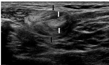
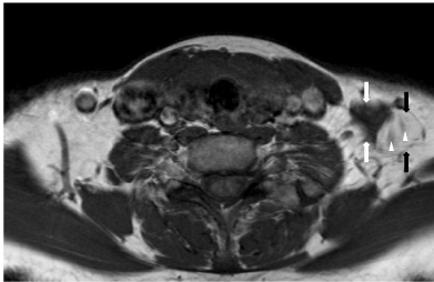
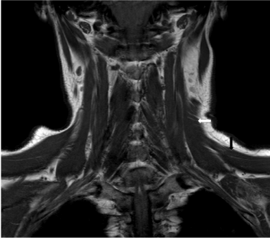
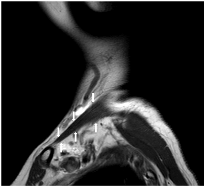
A 56 year old woman presented with a painless mass in the left posterior triangle for one month. Clinical examination revealed a solitary, firm, mobile, non-tender mass. This was suspected to be a pathologically enlarged lymph node and the patient underwent an ultrasound examination, which showed that the mass comprised two components; a superficial hyper-echogenic component with multiple linear striations resembling fat, and a deeper hypoechogenic component that was contiguous with a slender muscle of uncertain origin (Figure 1). Magnetic Resonance Imaging (MRI) of the neck was performed, which showed a unilateral muscle orientated obliquely in the posterior triangle with a lipomatous component interspersed eccentrically within the lateral aspect of the muscle belly (Figure 2). Superiorly, the muscle appeared as flat sheet merging with the upper fibers of the left trapezius muscle and attaching onto the external occipital protuberance, nuchal ligament and medial superior nuchal line (Figure 3a). Inferiorly, the muscle separated from the rest of the trapezius muscle, appearing as a slender muscle in the posterior triangle, and inserted onto the posterosuperior aspect of the left clavicle at the junction of its middle and medial third (Figure 3b). The imaging features were compatible with an anomalous left cleido-occipitalis cervicalis muscle with an intramuscular lipoma. As the patient had no discomfort and the lesion was acceptable cosmetically, this patient has been managed conservatively.
Figure 1: Oblique US image of the left posterior cervical triangle showing a mass comprising a superficial, oval hyper-echogenic structure (black arrows) that contains multiple linear striations similar to the subcutaneous fat. This is suggestive of a lipomatous lesion. The deeper margin of the mass is inseparable from a hypo-echogenic linear structure (white arrows) containing linear striations, which is suggestive of muscle. The precise nature of the muscular component was unclear. A similar muscle was not present in the right side of neck.
Figure 2: Axial T1W MRI image of the neck at the level of the thyroid gland showing a unilateral left posterior triangle mass. Its medial aspect comprises a triangular shaped T1W hypointense structure (white arrows) that is compatible with muscle tissue. Its lateral aspect comprises a rounded T1W hyperintense structure (black arrows) that has similar signal to the surrounding fat and is suggestive of a lipomatous component. A couple of muscle fibres are also present centrally within the lipomatous component (arrowheads).
Figure 3a: T1W MRI images of origin and insertion of the anomalous left neck muscle. Coronal image (a) showing its superior portion (white arrow) that fans out and merges with upper fibres of the left trapezius muscle (black arrow). Superomedially, it inserts into the left nuchal/occipital region (not shown).
Discussion
Cervical muscular anomalies present most commonly as a painless palpable neck mass, or rarely as a hard mass due to protruberance at the musculotendinous insertion onto the clavicle or as thoracic outlet syndrome due to neurovascular compression [6-8]. However, the majority of muscular anomalies are asymptomatic, of which a small proportion are detected incidentally during neck dissections or on cross sectional imaging examinations performed for other indications.
The clinical examination findings of anomalous neck muscles are non-specific and the differential diagnosis is wide. This includes cervical lymphadenopathy, thrombosed vein/varix, developmental or acquired neck cyst, hemangioma, lymphovenous vascular malformation, nerve sheath tumour (neurofibroma or schwannoma) or glomus tumour. Fortunately, ultrasound is excellent for differentiating the vast majority of neck masses, including by virtue of the typical sonographic appearances of different tissue constituents such as muscle and fat. Nevertheless, cross sectional imaging studies may be required for suspected muscular anomalies to determine the precise muscle involved. Besides an anomalous cleido-occipitalis muscle, other conditions of muscular origin can also involve the posterior triangle including a levator claviculae muscle, an accessory scalene muscle, or hypertrophy of the levator scapulae muscle (the latter may occur in response to spinal accessory nerve injury during neck dissection with denervation atrophy of the trapezius muscle) [3]. A levator claviculae muscle is worth highlighting as it is reportedly the commonest muscular anomaly to present as a neck mass. This accessory muscle arises from the upper cervical transverse processes and attaches to the clavicle in the middle or lateral third [7-11]. In this regard, it is also worth noting that anomalous muscles in the posterior triangle may superficially resemble enlarged lymph nodes on single axial sections of CT or MRI examinations. This pitfall can be avoided by careful inspection of all image sections.
The presence of a lipoma within a cleido-occipitalis muscle has not been reported previously. Although we cannot be certain, we postulate that their combined occurrence was co-incidental in view of the fact that cervical lipomas are very common. It also is probable that the lipomatous component had contributed to the overall bulk of the mass resulting in the clinical presentation of a palpable swelling. Most muscular anomalies can be managed conservatively although lesions causing symptomatic neurovascular compression may be excised [4]. We present this case to increase awareness of this uncommon condition, which has potential to cause diagnostic confusion both clinically and on imaging.
References
- O'Sullivan ST, Kay SP (1998) An unusual variant of the levator claviculae muscle encountered in exploration of the brachial plexus. J Hand Surg Br 23: 134-135.
- Wood J (1864) On some varieties in human mycology. Prod R Soc Lond 13: 299-303.
- Sarikcioglu L, Donmez BO, Ozkan O (2001) Cleidooccipital muscle: an anomalous muscle in the neck region. Folia Morphol (Warsz) 60: 347-349.
- Kwak HH, Kim HJ, Youn KH, Park HD, Chung IH (2003) An anatomic variation of the trapezius muscle in a Korean: the cleido-occipitalis cervicalis. Yonsei Med J 44: 1098-1100.
- Rahman HA, Yamadori T (1994) An anomalous cleido-occipitalis muscle. Acta Anat (Basel) 150: 156-158.
- Shaw AS, Connor SE (2004) Unilateral levator claviculae muscle mimicking cervical lymph node enlargement in a patient with ameloblastoma. Dentomaxillofac Radiol 33: 206-207.
- Hug U, Burg D, Meyer VE (2000) Cervical outlet syndrome due to an accessory part of the trapezius muscle in the posterior triangle of the neck. J Hand Surg Br 25: 311-313.
- Ruiz Santiago F, Lopez Milena G, Chamorro Santos C, Tristan Fernandez JM (2001) Levator claviculae muscle presenting as a hard clavicular mass: imaging study. Eur Radiol 11: 2561-2563.
- Fasel J, Gailloud P, Terrier F (1994) Three-dimensional reconstruction of a levator claviculae muscle. Surg Radiol Anat 16: 303-305.
- Loukas M, Sullivan A, Tubbs RS, Shoja MM (2008) Levator claviculae: a case report and review of the literature. Folia Morphol (Warsz) 67: 307-310.
- Rubinstein D, Escott EJ, Hendrick LL (1999) The prevalence and CT appearance of the levator claviculae muscle: a normal variant not to be mistaken for an abnormality. AJNR Am J Neuroradiol 20: 583-586.
Citation: Lee RKL, Bhatia KSS (2012) An Intramuscular Lipoma Developing within an Anomalous Cleido- Occipitalis Cervicalis Muscle. Otolaryngology 2:116. DOI: 10.4172/2161-119X.1000116
Copyright: © 2012 Lee RKL, et al. This is an open-access article distributed under the terms of the Creative Commons Attribution License, which permits unrestricted use, distribution, and reproduction in any medium, provided the original author and source are credited.
Share This Article
Recommended Journals
Open Access Journals
Article Tools
Article Usage
- Total views: 18018
- [From(publication date): 8-2012 - Apr 07, 2025]
- Breakdown by view type
- HTML page views: 13325
- PDF downloads: 4693


