Induction Therapy with S-1 for Spindle Cell Carcinoma of the External Auditory Canal
Received: 04-Oct-2013 / Accepted Date: 30-Oct-2013 / Published Date: 27-Nov-2013 DOI: 10.4172/2161-119X.1000149
Abstract
We report an unusual case of spindle cell carcinoma of the external auditory canal, which responded well to two weeks’ treatment with the oral fluoropyrimidine anticancer drug S-1 (80 mg/body/day) followed by surgical resection. After the S-1 treatment, the tumor was significantly reduced in size, and a pathological examination only detected small lesions composed of malignant cells. We then conducted a lateral temporal bone resection combined with left modified neck dissection. The patient did not demonstrate any evidence of local recurrence or distant metastasis during the 5-year follow-up period. It is suggested that S-1 played an important role in reducing the size of the tumor to allow radical resection of the tumor with a sufficient surgical margin in the present case and that induction chemotherapy with S-1 prior to surgery might be an effective treatment strategy for controlling spindle cell carcinoma of the ear.
Keywords: Spindle cell carcinoma, External auditory canal, S-1
255384Introduction
Spindle Cell Carcinoma (SpCC) is an unusual pathological variant of Squamous Cell Carcinoma (SCC), which is the most common neoplasm in the head and neck region. SpCC is more frequently observed in the larynx and has been studied in detail [1-4]. In the larynx, SpCC constitutes 1-2 % of all SCC, and it is more predominant in males than in females [2,5]. The age prevalence widely ranges from 20 to 90 years, with the highest in the 50-60-year age group. Similar to SCC, cigarette smoking and alcohol consumption are associated with the development of SpCC [1,2,5]. SpCC is characterized by a gross polypoid or ulcerated configuration [1,2]. Surgery is considered to be used as a first-line treatment since it is also conducted to treat conventional SCC [1,2]. However, prognosis on survival, which is affected by recurrences and metastases, is likely to be poorer in SpCC than in most conventional SCC [1,2,6].
On the other hands, only one case of SpCC of the External Auditory Canal (EAC) has been reported [7]. Therefore, optimal therapeutic strategy is yet to be determined, although in cases of SCC of EAC, en bloc resection involving a sufficient tumor-free surgical margin has been reported to be important for patient survival [8].
We report an extremely rare case of SpCC of EAC, which responded well to treatment with the oral fluoropyrimidine anticancer drug S-1 (tegafur, gimeracil, and oteracil) followed by surgical resection of the tumor with a sufficient surgical margin.
Case Report
In 2008, a 75-year-old female presented with a gradually growing mass and effusion in her left ear two months before she was admitted to hospital. She had suffered from recurrent episodes of bilateral chronic otitis media since childhood, but had no history of smoking, alcohol consumption, or radiation exposure.
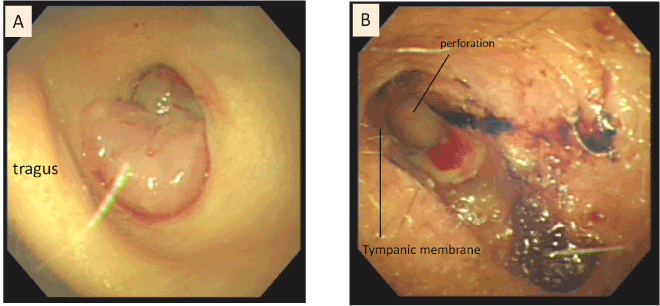
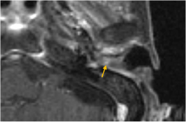
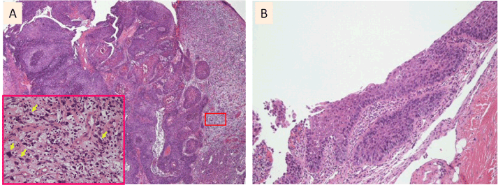
A flexible endoscopic examination revealed a bleeding polypoid tumor that had completely occluded the left EAC (Figure 1A); thus, it was not possible to perform a thorough evaluation of the ear canal and tympanic membrane behind the tumor. However, it was evident that the tumor was not adherent to the surrounding tissue. Contrastenhanced Computed Tomography (CT) of the temporal bone (Figure 2) showed a 2 cm mass located in the left EAC together with minor bony erosion of the anterior wall. A small amount of soft tissue, which was considered to be related to the patient’s previous chronic otitis media, was observed in the left middle ear cavity. Magnetic Resonance Imaging (MRI) (Figure 3) demonstrated that an enhancement of the isolated mass in the left canal had extended up to the tympanic membrane medially, but had not invaded into the middle ear. There was no evidence of cartilage invasion underlying the external ear canal. A pathological examination of a 2-mm biopsy sample taken from the tumor revealed the coexistence of a well-differentiated SCC component and a spindle cell component. There was no evidence of transition between the two components (Figure 4A). We diagnosed the patient with SpCC of the EAC.
Figure 1: Endoscopic views of the lesion in the left external auditory canal before (A) and after (B) induction chemotherapy involving the administration of S-1 for two weeks followed by surgery. A: An easily bleeding polypoid tumor had completely occluded the left EAC. B: After the induction chemotherapy, the tumor had markedly decreased in size, resulting in the erosion of the skin in the EAC.
Figure 2: A: Preoperative axial CT of the temporal bone showing an isolated tumor in the left external ear canal together with minor bony erosion of the anterior wall of the canal (arrow). B: Preoperative coronal CT revealing a soft tissue mass in the left external ear as well as the attic of the tympanic cavity. It was unclear whether the tumor extended into the tympanic cavity.
Figure 3: Preoperative axial MRI demonstrating an enhancement of the mass localized in the left external auditory canal. The tumor extended up to the tympanic membrane medially, but there was no evidence of middle ear invasion. The tumor had not invaded the cartilage underlying the external ear canal.
Figure 4: Pathological examination before (A) and after (B) induction chemotherapy involving the administration of S-1 for two weeks followed by surgery. A: The tumor displayed a biphasic appearance involving the coexistence of a spindle cell component (right side) and a squamous cell carcinoma component (left side). The inset shows a magnified image of the pleomorphic spindle cells (arrow) in the boxed region. B: A pathological examination of the surgical specimen revealed carcinoma-in-situ without invasion into the surrounding tissues.
Since the imaging studies demonstrated bony erosion of the ear canal where the tumor was limited, we suggested S-1 chemotherapy (Taiho, Tokyo, Japan) in addition to en bloc resection of the temporal bone. The administration of S-1 for two weeks resulted in a significant reduction in the size of the tumor (Figure 1B). We then performed a lateral temporal bone resection combined with left modified neck dissection and removal of the superficial part of the parotid gland. A pathological examination of the surgical specimen revealed localized intraepithelial neoplasia in the external ear canal (Figure 4B) without any metastatic lymph nodes. The patient’s clinical course was uneventful during the 5-year follow-up period, and no evidence of local recurrence or distant metastasis was observed.
This study was approved by the clinical research ethics board of Nara Medical University Hospital (Approval No.669).
Discussion
SpCC is an unusual variant of poorly differentiated squamous cell carcinoma consisting of spindles of epithelial cells that histologically resembles sarcoma. It most commonly occurs in the upper aerodigestive tract, e.g., in the larynx [1-4], nasal cavity [9], or hypopharynx [2]; however, SpCC of the external auditory canal is extremely rare. Although the optimal treatment strategy for SpCC has not been determined, surgery is considered to be the first-line treatment since it is also used to treat conventional SCC [1,2]. SpCC is likely to manifest more aggressive behavior than most conventional SCC [1,2,6]. In the present case, imaging studies demonstrated that a tumor mass located in EAC extending up to the tympanic membrane with bony erosion of the anterior wall. Therefore, we treated the present patient with S-1 as an induction therapy in order to allow radical resection of the tumor with a sufficient surgical margin. This is the first report to describe the treatment of SpCC of the ear with S-1.
S-1 is an oral antitumor agent that consists of tegafur (FT), a prodrug of 5-FU and two modulators, 5-chloro-2, 4-dehydroxypyimidine and potassium oxonate. This drug was designed to enhance the efficacy of 5-FU by maintaining therapeutic serum 5-FU concentrations for long periods of time. In addition, it also reduces adverse reactions to FT in the gastrointestinal mucosa [10,11]. S-1 has been demonstrated to have antitumor effects in patients with various malignant diseases, including head and neck cancer [12]. There have been several reports about the administration of S-1 as adjuvant chemotherapy for head and neck cancer. One report recommended that S-1 should be administered at a dose of 60 mg/m2/day for 5 days, followed by 2 days off, in combination with irradiation (total dose: 64-70 Gy) for 6-7 weeks [12], while another study recommended that S-1 should be administered every day at a dose of 80-100 mg/body/day for 2 weeks and then suspended for 1-2 weeks to allow radiotherapy to be performed (total dose: 60-70.2 Gy) [13,14]. On the basis of the results of these studies, we chose a treatment regimen in which S-1 was delivered at a dose of 80 mg/body/day every day for 2 weeks before surgery. This regimen produced good results without serious adverse events, suggesting that SpCC responds well to S-1. Although the prognosis of SpCC in head and neck region has been described to be unfavorable in some cases [1,3,15], the present case did not demonstrate any evidence of local recurrence or distant metastasis during the 5-year follow-up period.
At present, the treatment strategy for SpCC of the ear canal is not precisely specified since the only one case of SpCC was previously reported in the EAC [7]. Thus, it is necessary to identify the clinicopathological characteristics of SpCC of the external auditory canal and gather data from patients and case studies in order to elucidate the optimal treatment strategy for the disease. In this case, as the administration of S-1 produced a good response, S-1 chemotherapy prior to surgical resection could be a useful and practical treatment for SpCC of the ear canal.
Conclusion
The favorable results obtained in the current case suggest that induction chemotherapy with S-1 prior to surgery is a feasible way of achieving control over spindle cell carcinoma of the external auditory canal.
References
- Thompson LD, Wieneke JA, Miettinen M, Heffner DK (2002) Spindle cell (sarcomatoid) carcinomas of the larynx: a clinicopathologic study of 187 cases. Am J Surg Pathol 26: 153-170.
- Olsen KD, Lewis JE, Suman VJ (1997) Fuziform cell carcinoma of the larynx and hypopharynx. Otolaryngol Head Neck Surg 116: 47-52.
- Lewis JE, Olsen KD, Sebo TJ (1997) Spindle cell carcinoma of the larynx: review of 26 cases including DNA content and immunohistochemistry. Hum Pathol 28: 664-673.
- Miyahara H, Tsuruta Y, Yane K, Ogawa Y (2004) Spindle cell carcinoma of the larynx. Auris Nasus Larynx 31: 177-182.
- Viswanathan S, Rahman K, Pallavi S, Sachin J, Patil A, et al. (2010) Sarcomatoid (Spindle Cell) Carcinoma of the Head and Neck Mucosal Region: A Clinicopathologic Review of 103 Cases from a Tertiary Referral Cancer Centre. Head Neck Pathol 4: 265-275.
- Batsakis JG, Suarez P (2000) Sarcomatoid carcinomas of upper aerodigestive tracts. Adv Anat Pathol 7:282-293.
- Quaranta A, Berardi P, Piscitelli D, Fiore MG, Calace A, et al. (2007) Spindle cell carcinoma of the external auditory meatus with intracranial extension: histological, immunohistochemical and electron microscopic evaluation. Acta Otolaryngol 127: 105-109.
- Nakagawa T, Kumamoto Y, Natori Y, Shiratsuchi H, Toh S, et al. (2006) Squamous cell carcinoma of the external auditory canal and middle ear: an operation combined with preoperative chemoradiotherapy and a free surgical margin. Otol Neurotol 27:242-248.
- Terada T, Kawasaki T (2011) Spindle cell carcinoma of nasal cavity. Int J Clin Oncol 16: 165-168.
- Shirasaka T, Nakano K, Takechi T, Satake H, Uchida J, et al. (1996) Antitumor activity of 1 M tegafur-0.4 M 5-chloro-2,4-dihydroxypyridine-1 M potassium oxonate (S-1) against human colon carcinoma orthotopically implanted into nude rats. Cancer Res 56:2602-2606.
- Fukushima M, Satake H, Uchida J, Shimamoto Y, Kato T, et al. (1998) Preclinical antitumor efficacy of S-1: a new oral formulation of 5-fluorouracil on human tumor xenografts. Int J Oncol 13: 693–698.
- Nakata K, Sakata KI, Someya M, Miura K, Hayashi J, et al. (2013) Phase I study of oral S-1 and concurrent radiotherapy in patients with head and neck cancer. J Radiat Res 54:670-683.
- Tsuji H, Nagata M, Inoue T, Minami T, Iwai H, et al. (2006) Clinical Phase I trial of concurrent chemo-radiotherapy with S-1 for T2N0 glottic carcinoma. Jpn J Cancer Chemother 33 Suppl 1:163-166.
- Tsukuda M, Ishitoya J, Mikami Y, Matsuda H, Horiuchi C, et al. (2009) Analysis of feasibility and toxicity of concurrent chemoradiotherapy with S-1 for locally advanced squamous cell carcinoma of the head and neck in elderly cases and/or cases with comorbidity. Cancer Chemo Pharm 64: 945-952.
- Leonardi E, Dalri P, Pusiol T, Valdagni R, Piscioli F (1986) Spindle cell squamous carcinoma of head and neck region; a clinicopathologic and immunohistochemical study of eight cases. ORL J Otorhinolaryngol Relat Spec 48: 275-281.
Citation: Yamanaka T, Shimizu N, Enomoto Y, Hosoi H (2013) Induction Therapy with S-1 for Spindle Cell Carcinoma of the External Auditory Canal. Otolaryngology 3:149. DOI: 10.4172/2161-119X.1000149
Copyright: © 2013 Yamanaka T, et al. This is an open-access article distributed under the terms of the Creative Commons Attribution License, which permits unrestricted use, distribution, and reproduction in any medium, provided the original author and source are credited.
Share This Article
Recommended Journals
Open Access Journals
Article Tools
Article Usage
- Total views: 15110
- [From(publication date): 11-2013 - Nov 24, 2024]
- Breakdown by view type
- HTML page views: 10600
- PDF downloads: 4510


