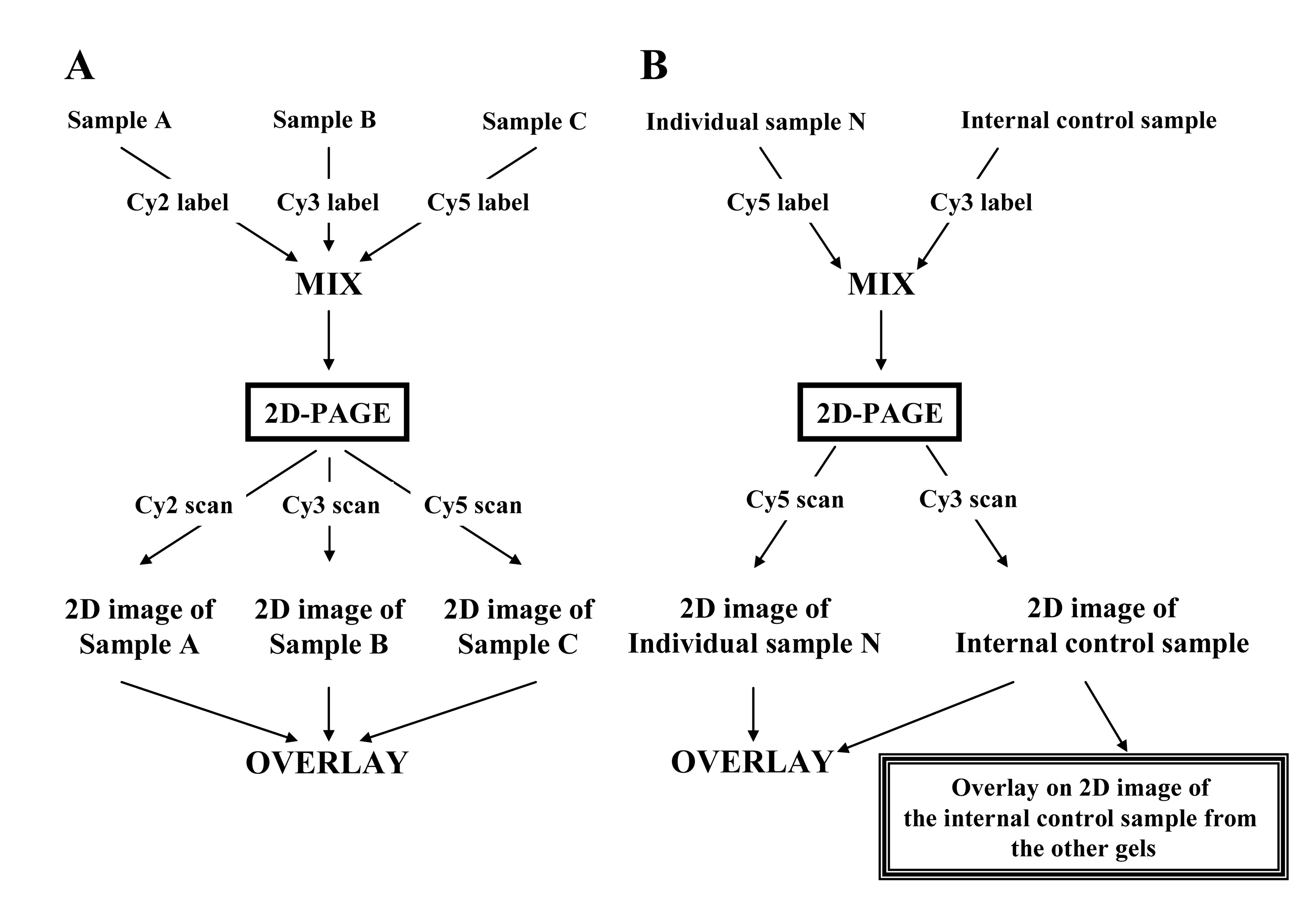
Figure 3: Methodology employed for 2D-DIGE experiments using multiple samples. A. Three samples are labeled with Cy2, Cy3, and Cy5 respectively, mixed together, then separated by 2D-PAGE. Laser scanning for each sample generates the 2D images, which are then overlaid so that they can be examined comparatively. B. The individual sample and the internal control sample are labeled with Cy5 and Cy3 respectively, mixed together and separated by 2D-PAGE. The same procedure is repeated for all individual samples. The Cy3 images of the internal control sample in the different gels are compared to normalize gel-to-gel variations. The ratio of the Cy5 and Cy3 spot intensity in the same gels is considered as the normalized intensity of the protein spots.