Ischaemic Optic Neuropathy - Arteritic
Received: 04-Apr-2016 / Accepted Date: 01-Jun-2016 / Published Date: 04-Jun-2016 DOI: 10.4172/2476-2075.1000116
Keywords: Ischaemic optic neuropathy; Orbital optic nerve; Vision
6609Introduction
Ischaemic optic neuropathy (ION) is the commonest adult optic nerve disorder encountered worldwide and can be expected to increase in incidence in our ageing population. The condition has been classified as a) anterior (AION) affecting the optic nerve head and b) posterior (PION) involving that portion of the optic nerve behind its immediate retrolaminar portion. Furthermore there are two pathological varieties of the disease c) Arteritic (AAION) almost exclusively associated with Giant Cell Arteritis (GCA) and d) Nonarteritic (NA-AION or less correctly NAION) usually associated with diabetes, hypertension and hypercholesterolaemia. A recent treatise on the subject [1] runs to more than 600 pages.
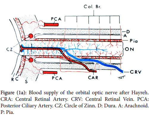
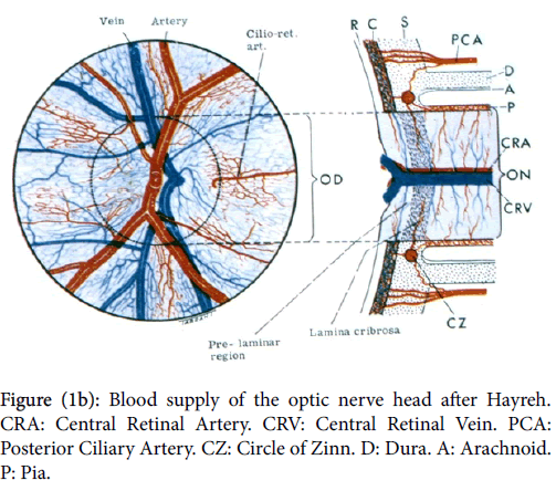
In order to understand the pathology of ION knowledge of the complex vascular anatomy of the optic nerve head (ONH) and its more posterior part is required. This was not clarified until the mid 1960’s by the work again of Hayreh [2], when he showed that the ONH is supplied in the main by the ciliary vascular system and not by the ophthalmoscopically visible central retinal artery; furthermore the more posterior part of the nerve is supplied from its surrounding pial plexus fed from adjacent orbital branches of the ophthalmic artery (Figure 1a).
The ophthalmic artery is the first intracranial branch of the internal carotid when it emerges from the cavernous sinus. The central retinal artery, also a branch of the ophthalmic, only supplies the surface/ retinal layer of the ONH (Figure 1b) before it proceeds to supply the inner layers of the retina.
Arteritic ION
GCA is by far the commonest condition causing arteritic ischaemic optic neuropathy which is almost exclusively of the anterior variety i.e AAION and is a common cause of blindness in old age. It is of particular importance because the diagnosis is often missed when the first eye is involved and only diagnosed when the second is affected and the patient blind. Unfortunately recovery of vision is not to be expected and the condition is one of the commonest eye diseases leading to litigation for failure of diagnosis.
Variable figures for the incidence of GCA have been reported the condition being much commoner in the northern hemisphere. A 1979 report [3] from the Lothian region Scotland (population then circa 750,000) gave an incidence of 1.3 cases per 100,000 populations per annum and 4.23/100,000/annum over the age of fifty. A survey from the same area, now with 800K population, in 2010 to 2015 [4] showed a fourfold yearly increase in cases, thus from expecting to encounter less than one case per month in 1979 now they will see one new case nearly every week.
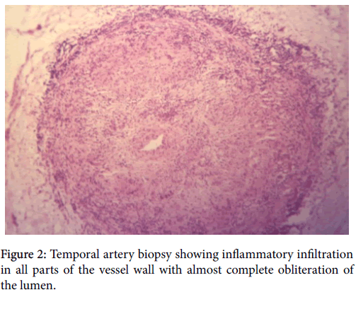
GCA is a disease of large and medium sized blood vessels and most commonly involves the external carotid artery and its branches giving rise to the classical symptoms of pain and tenderness in the area supplied especially the temporal region of the head thus the original name for the condition Temporal Arteritis. It has been realised for many years that visual and ocular complications occur in at least 50% of cases of this classical arteritis, the commonest complication being visual loss from optic nerve ischaemia often preceded by transient visual obscurations (TIA’s). Conversely the optic nerve blood supply is from the internal carotid system via branches of the ophthalmic artery in particular the ciliary vessels which supply the ONH. Strangely these are the only branches of the internal carotid involved in GCA and it is notable that its branches supplying the brain are not affected [5]; this anomaly has yet to be explained. The ischaemic process is due to gradual occlusion of the ON vasculature from a progressive inflammatory process as seen in the biopsy specimens in Figure 2.
GCA appears in two forms classical and occult [6]. The former presenting with pain and tenderness in the temple and in other parts supplied by the external carotid, whereas the occult and more dangerous form presents with acute and extreme visual loss usually due to an AAION and in 10% of cases with a central retinal artery occlusion. In this variety there may be no general symptoms or abnormal signs in the temples but a temporal artery biopsy (TAB) will be positive. In both varieties the ESR and CRP are raised and unless diagnosed and treated at the initial presentation the second eye will be similarly involved within one day in one third of patients, and within one week in a further third [3]. GCA is sometimes associated with another medical condition polymyalgia rheumatica.
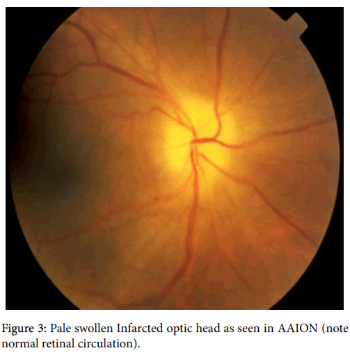
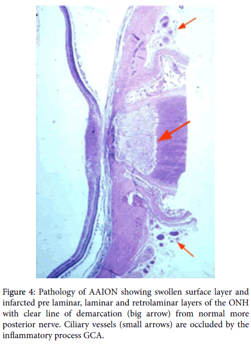
The clinical picture of AAION is one of acute infarction of the ONH with extreme or complete loss of vision, a marked relative afferent pupil defect (RAPD) or complete loss of the pupillary reactions to light if both eyes are involved. The nerve is pale, swollen and infarcted as seen in Figure 3. The pathology is shown in Figure 4 with destruction of the prelaminar, laminar and retro-laminar layers of the ONH all supplied by the ciliary vessels which are occluded by the inflammatory process. Clearly recovery of vision due to such a pathological process cannot be expected. In many cases the ONH becomes cupped later and be mistaken for advanced glaucoma.
Treatment of AAION is usually designed to prevent involvement of the other eye with immediate corticosteroids monitored by regular ESR/CRP readings, and in many cases long term treatment is required [7]. Classical GCA is treated in the same way for a minimum period of 2 years and treatment in all cases can only be stopped when the ESR/CRP remains normal on attempted withdrawal.
Recurrence can occur and has been reported as long as 7 years from diagnosis [8]. A TAB is essential in establishing the diagnosis and should be performed as soon as possible and at least within forty eight hours of the patient’s presentation. Follow-up is preferable in the Eye clinic and an up to date ESR /CRP result should be available at the time of consultation.
Posterior ischaemic optic neuropathy (PION) has been reported rarely in association with GCA [9] and is due to involvement of the posterior optic nerve blood supply. The patient presents with a central visual defect and the optic nerve remains normal for up to six weeks similar to the situation in retro-bulbar optic neuritis. PION is a diagnosis of exclusion so that a compressive cause must be investigated.
Other aetiologies for arteritic AION and PION are rare and have not been confirmed histologically. Systemic Lupus and polyarteritis nodosa are two possible causes along with and other rarer systemic vasculitides.
References
- Hayreh SS (1969) Blood supply of the optic disc and its role in optic atrophy, glaucoma and oedema of the optic disc. Brit J Ophthalmol53: 721-748.
- Jonasson F, Cullen JF, Elton RA (1979) Temporal Arteritis –A 14 year epidemiological, clinical and prognostic study. Scot Med J 24:111-117.
- Cullen JF, Thavaratnam LK (2016) Neuro-ophthalmic disease patterns in Southeast Asia with particular reference to giant cell arteritis. Eye News 22:26-29.
- Butt Z, Cullen JF, Mutlukan E (1991) Pattern of arterial involvement of the head, neck and eyes in giant cell arteritis. Brit J Ophthalmol 75: 368-371.
- Cullen JF (1967) Occult Temporal Arteritis: A common cause of blindness in old age. Brit J Opthalmol51:513-525.
- Culen JF, Chan J, Wong CF, Chew WC (2010) Giant Cell (Temporal) Arteritis in Singapore,an Occult Case and the Rationale of Management. Singapore Med J 51:73-77.
- Cullen JF (1972) Temporal Arteritis: Occurence of Ocular Complications seven years following Diagnosis. Brit J Ophthalmol 56:584-588.
- HayrehSS (2004) Posterior Ischaemic Optic Neuropathy: Clinical features pathogenesis and management. Eye 18:1188-1206.
Citation: Cullen JF (2016) Ischaemic Optic Neuropathy-Arteritic. Optom Open Access 1:116. DOI: 10.4172/2476-2075.1000116
Copyright: © 2016 Cullen JF. This is an open-access article distributed under the terms of the Creative Commons Attribution License, which permits unrestricted use, distribution, and reproduction in any medium, provided the original author and source are credited
Share This Article
Recommended Journals
Open Access Journals
Article Tools
Article Usage
- Total views: 12699
- [From(publication date): 6-2016 - Nov 23, 2024]
- Breakdown by view type
- HTML page views: 11910
- PDF downloads: 789

